


4. Gravitropism as a morphogenetic model
My interests in gravitational biology arose from one of those 'twists of fate' stories. Early in the afternoon of 26 June 1989 I received a telephone call from Dr Greg Briarty of the University of Nottingham asking if I might be interested in suggesting mycological projects for inclusion in the Juno space mission. At the time, I had already developed interests in mushroom (especially Coprinopsis) developmental biology, but had not thought very seriously about the part that gravity might play in cell biology or development. The phone call spurred that interest and I was quickly able to assemble a proposal.
My proposal was accepted as one of the 26 experiments to be done on the Juno flight which was planned for launch from the Soviet space centre in Spring 1991, timed to commemorate Yuri Gagarin’s triumphant first step. On 12 April 1961 Yuri Gagarin, the first human to venture beyond the pull of Earth’s gravity, was launched in Vostok 1 for an orbital flight that lasted just 108 minutes; but when he unstrapped his parachute after landing he had proved that man could survive the rigours of a rocket launch, could live and work in orbit, and be returned to ground safe and well (despite medical predictions of the time that, removed from hydrostatic restraint of gravity, body fluids would flood into his chest and stop the beating of his heart and inflation of his lungs!). From the very beginning the Juno mission was planned to provide British scientists with the opportunity to fly experiments exploiting the virtual absence of gravity on board an orbiting spacecraft, funded by British and international companies with UK and Soviet links (Moore, 1990; Moore, 1991a).
Helen Sharman eventually flew the eight-day mission to the Soviet orbital complex Mir in 1991, but by that time the scientific hopes of the Juno Mission had been dashed by lack of sponsorship funding. Unfortunately for Juno, like other attempts to put British science into orbit, it felt the kiss of death of a 1989 report to the ruling Science and Engineering Research Council which concluded that there is 'no strong case for becoming seriously involved [in microgravity research].'
British fungi stayed on the ground, but they did contribute to a healthy amount of research on gravitropism. As part of the preparation for my Juno proposal I conducted a full review of the more than 100 year history of research on gravitropism in fungi (Moore, 1991b). This review made it clear that ‘mushroom’ fruit bodies (polypore and agaric alike) exhibit a number of tropisms of which anemotropism, gravitropism, phototropism and thigmotropism have been clearly demonstrated. At any one time, one tropism usually predominates but the inferior tropisms can be demonstrated if the predominating ones can be removed by manipulation of the growth conditions. In ascending order of priority, the hierarchy appears to be: thigmotropism (touch), gravitropism, anemotropism (air currents), phototropism (light). During the course of development of a fruit body different tropisms predominate at different times. The youngest fruit body initials grow perpendicularly away from their substratum. The nature of this tropism is completely unknown but could be a chemotropism to moisture. Perpendicular growth of fruit body initials has been remarked upon in experiments at a variety of light intensities and in gravitational fields from ± 0 to 4.5 g. The fruit-body primordium then becomes first positively phototropic, but later negative gravitropism predominates (Moore, 1991b).
The switch between predominance of the two tropisms has been associated with the onset of sporulation in a number of different studies. The major adjustment of the direction of growth in response to a tropic stimulus is made by the mushroom stem. It is the apex of the stem which makes the most immediate gravitropic response. Gravitropic growth curvatures are limited to the normal growth zones of the stem and seem to depend on re-allocation of available growth resources. If the fruit body is reoriented late in the growth of the stem, it may not be able to respond fully. In these cases gravitropic movements of the cap may still be able to bring the hymenophore back to the vertical. And this is the crucial point: the hymenophore, whether made up of gills or pores, must be vertical to allow basidiospores to fall freely under the influence of gravity alone, either in the space between adjacent gills in 'agarics' or down the centre of the tubes that make up poroid fruit bodies.
Mechanical forces may influence and contribute to the ‘gravitropic’ response (the stem being put under mechanical leverage by the mass of the cap at its apex) but this has not been experimentally examined. The hymenophore (gill, tube or tooth) is positively gravitropic and responds independently of the stem. Bracket polypores do not show tropisms but exhibit gravimorphogenetic responses such that gross disturbance leads to renewal of growth to produce an entirely new fruiting structure suitably reoriented to the new spatial position. One experiment performed on an orbiting space station suggests that, in the absence of a light stimulus, gravity may be required for initiation of fruiting in Polyporus brumalis. Otherwise, the indications from both clinostat and space-borne experiments are that the basic form of the mushroom (overall tissue arrangement of stem, cap, gills, hymenium, veil) in agaric and polypore alike is established independently of the gravity vector (Moore, 1991b).
Abnormal stem growth has been observed in clinostat cultures of Lentinus tigrinus and Polyporus brumalis, but the morphogenetic event which seems most dependent on gravity is sporulation (in the broadest sense). Cultures of P. brumalis on orbiting space craft fail to produce the poroid hymenophore and in clinostat experiments on the ground even karyogamy was rare in similar cultures. Coprinopsis cinerea grown on the clinostat was able to produce apparently normal fruit body primordia which failed to produce spores and then aborted, forming a new flush of primordia on the old. Taken together with the clear association between observation of gravitropism and the onset of sporulation, the implication is that commitment to the meiosis-sporulation pathway both requires the gravity vector and couples it in some way to fruit-body growth.
The earlier literature offers no convincing evidence for a graviperception mechanism in fungi. There is no evidence for any organised means of communicating the gravitropic stimulus once it has been perceived. Indeed, most early authors were convinced that the apparently coordinated expression of gravitropic response is in truth a common, but independent, response by the individual component hyphae of the structure concerned. This may well be preconditioned by work on tropisms using Phycomyces sporangiophores which are single, independent, hyphae; but an echo of this conviction returned very much later when our work on computer simulations of fungal growth showed that multihyphal morphologies similar to differentiated fungal fruit bodies could be generated by simulations in which all the cyberhyphae in the computer model were reacting to the same set of instructions (Meškauskas, McNulty & Moore, 2004; [view section now]).
For the negatively gravitropic Phycomyces sporangiophores there is evidence that the vacuole floats in the protoplasm. It is thought that this could affect protoplasmic volumes above and below the vacuole in a horizontally-placed sporangiophore such that a greater proportion of the cell's potential for wall growth was adjacent to the lower wall, enabling that wall to extend more than the upper and so curve the sporangiophore upwards. This is not only an attractive way of accounting for the asymmetric wall growth of negative gravitropism, but since the relative density of the vacuole can presumably be controlled by regulation of water influx and efflux, it is also an attractive means of accounting for the control of gravitropic responses. Phycomyces also exhibits a response to the mechanical consequences of reorientation which is additional to (and different from) the longer term gravitropic response. If this also applies in higher fungi, it is possible that some of the apparent organisation in the response of independent hyphae across a structure like a fruit-body stem of hymenophore could be a concerted response to altered distribution of mechanical loads rather than coordination of response by some morphogen or hormone chemical (Moore, 1991b).
 |
| Fig. 22. Gravitropism of Agaricus bisporus: an inverted fruit body is able to regain the vertical. Cutting the fruit body down the centre shows that this has been achieved at the expense of other aspects of fruit body morphogenesis, like the trapped and poorly-developed gills on the inside of the curved stem and the structural disruption of the stem itself. Although my own research efforts have been devoted to Coprinopsis, this image shows just how important it is for a mushroom to be vertical. It's important enough to rip the structure apart to achieve; it's important enough to sacrifice half of the mushroom to achieve. Experiment carried out and photographed by Dr Halit Umar. |
Fig. 22 shows an Agaricus bisporus fruit body which has restored itself to the vertical after being inverted. The photograph demonstrates that the gravitropic reaction is an absolute imperative; evidently, development of the cap on the inside of the curved stem has been aborted to enable the stem to bend sufficiently, and the bending has pulled apart the structure of the stem. Such radical re-engineering of fruit body structure is permitted because vertical orientation of the fruit body is essential for effective spore release and it may be achieved even at the expense of other aspects of fruit body morphogenesis (Moore, 1991b). This image of A. bisporus gravitropism dramatises the process but this is a difficult organism to work with routinely and our work has concentrated on Coprinopsis cinerea gravitropism. This, together with research by our colleagues on Flammulina velutipes gravitropism, was the subject of a Centenary Review (Moore, Hock, Greening, Kern, Novak Frazer & Monzer, 1996) which I do not intend to repeat here. I do, however, want to emphasise a few points about our experiments.
One of the first things we set out to establish was the exact kinetics and mechanics of gravitropism in Coprinopsis cinerea (Kher, Greening, Hatton, Novak Frazer & Moore, 1992). We developed a standardised, simple experiment in which first complete fruit bodies and eventually fruit body stems with the caps removed were placed horizontally (fixed with a mapping pin through their basal bulb) on a support in an environmental chamber to be observed continuously using video recordings. We found that the stem became gravireceptive after the cap had completed meiosis, beginning to bend within 30 minutes of being placed horizontal. Stem bending first occurred in the apical 15% of its length, and then the position of the bend moved rapidly towards the base, traversing 40% of stem length in 2.5 h. Meanwhile, the stem elongated by 25%, mostly in its upper half but also in basal regions. If the apex was pinned in the horizontal position the stem base was elevated but overshot the vertical, often curling through more than 300°. When the base was pinned to the horizontal (considered analogous to the normal situation), 90% of the initial bend was compensated as the stem brought its apex accurately upright, rarely overshooting the vertical. The apex had to be free to move for this curvature compensation to occur.
 |
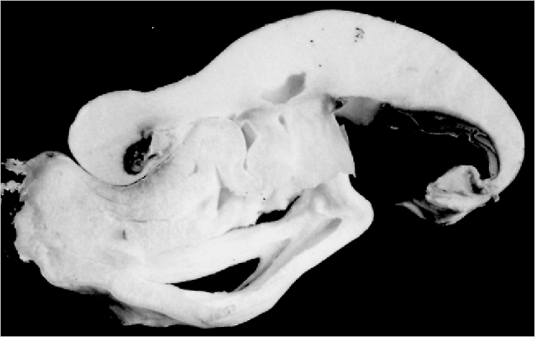
| Fig. 23. Example of a clinostat experiment showing the time course of the reaction of a Coprinopsis cinerea stem to 45 minutes horizontal gravistimulation. A 60 mm long stem was mounted horizontally, with cap removed, on a type 2 clinostat (see Fig. 24) and held stationary for 45 minutes and then the clinostat was switched on to rotate the specimen around its long axis at 2 rpm. This rotation randomises the gravity vector and effectively terminates the gravistimulation. Video recording maintained a continuous observation of the clinostat from the time of mounting the specimen (time zero); the camera operated in red light to avoid complicating stimulation of the blue-light photoreceptor known to be present in fruit bodies. Measurements were made from the video recordings at the end of the experiment. The clinostat was turned off after 180 minutes but video observation continued for a further 30 minutes. The plots show the overall length of the specimen (circles) and the angle of the stem tip to the (horizontal) axis of rotation (squares). An important observation is that the stem elongated throughout the experiment at a uniform rate; extending from 60 to 93 mm in about 3 h. This stem was fixed on the rotational axis of the stationary clinostat (and thus horizontal) for 45 min before rotation was started. Since the reaction time is less than 30 min this stem began to bend before clinostating, reaching an angle of 36° to the rotational axis before the clinostat was switched on. After the start of clinostating it bent further, reaching an angle of 48° but then, in the absence of further unidirectional gravistimulation the tip angle declined to 9° when the clinostat was switched off 180 min after the start of the experiment. Continued observation of the stationary (horizontal) stem showed that it immediately expressed negative gravitropism, reaching an angle of 72° to the horizontal within 30 min of the clinostat being stopped. These observations are representative of many others. Generalising: (a) the degree of bending achieved by the stem depended on the ‘dose’ of gravistimulation; (b) most stems reached a maximum angle of bend and then relaxed back towards the rotational axis (their original horizontal); and (c) stopping the clinostat resulted in immediate expression of the usual negative gravitropic response. Evidently, the gravitropic reaction requires continued stimulation for completion but sensitivity does not decline with increasing length. Modified after Hatton & Moore, 1992. |
Stems transferred to a clinostat (see below [view now]) after some minutes gravistimulation showed curvature which increased with the length of initial gravistimulation, indicating that continued exposure to the unilateral gravity vector was necessary for continued bending (Fig. 23). Such gravistimulated stems which bent on the clinostat subsequently relaxed back towards their original orientation. Reaction kinetics were unaffected by submergence in water, suggesting that mechanical events do not contribute, but submerged stems bent first at the base rather than apex. In air, the gravitropic bend appeared first near the apex and then moved towards the base, suggesting basipetal movement of a signal. In water, the pattern of initial bending was changed (from apex to base) without effect on kinetics. Taken together we think these results suggest that bending is induced by a diffusing chemical growth factor (whose extracellular propagation is enhanced under water) which emanates from the apical zone of the stem. The apex is also responsible for regulating compensation of the bend so as to bring the tip to the vertical. The nature of this latter stimulus is unknown but it is polarised (the apex must be free to move for the compensation to occur) and it may not require reference to the unilateral gravity vector (Kher, Greening, Hatton, Novak Frazer & Moore, 1992).
Importantly, we showed that mechanical stress was not involved in regulating stem gravitropism in Coprinopsis cinerea (Greening, Holden & Moore, 1993). We found we could remove large segments of the apical part of the C. cinerea stem (extending to about half its length) without affecting either the ability of the remaining stem to show gravitropic bending or its ability to compensate the curvature so induced and adjust to the vertical. However, gravitropic reaction time increased in direct proportion to the amount of stem removed. Application of lateral loads of up to 20 g had no adverse effects on adjustment of the stem to the vertical and the continuation of its vertical growth (the range of the fresh weights of the caps removed from the experimental stems was only 1 to 2 g). We concluded that sensing the distribution of extracellular mass and/or mechanical stress is unlikely to be a component of the control of gravitropic bending in C. cinerea stems. In particular, these experiments seem to exclude the possibility that the pendulum effect of the comparatively massive cap has any influence on recovery of the stem from a horizontal placement.
Using the standard gravitropism assay we found that inhibitors of Ca2+ transport and calmodulin-mediated Ca2+ uptake had no effect on gravity perception but did significantly diminish gravitropism. We used a Ca2+ channel blocker (verapamil); a Ca2+ ionophore (A23187); a Ca2+ chelator (BAPTA); and calmidazolium, an inhibitor of calmodulin-mediated Ca2+ uptake; using concentrations and treatments known to eliminate gravitropism and other tropisms in plant organs. We concluded that, under the conditions tested, Ca2+ is not involved in gravity perception by Coprinopsis stems, but does contribute to transduction of the gravitropic impulse. The results would be consistent with regulation of the gravitropic bending process requiring accumulation of Ca2+ within a membrane-bound compartment (Novak Frazer & Moore, 1993). Treatment of stems with an actin inhibitor caused a significantly delayed response, a result not observed with the Ca2+ inhibitors. This suggests that cytoskeletal elements may be involved directly in perception of gravity by Coprinopsis stems while Ca2+-mediated signal transduction may be involved in directing growth differentials that generate the gravitropic response (Novak Frazer & Moore, 1996). Interestingly, verapamil caused both diminished gravitropism and decreased growth of the stem. On the other hand, the ionophore, A23187, enhanced stem extension growth rate (by 30%) but decreased (by 43 %) the rate of response (that is, the rate of bending), suggesting that tropic bending may not result from a simple redistribution of the normal growth potential of the stem.
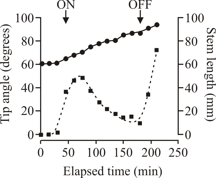
We built some clinostats to examine their influence on Coprinopsis stem gravitropism. A clinostat provides circular rotation at uniform speed around the horizontal axis. This does not remove the subject from the effects of gravity but a test subject mounted on the clinostat experiences altered vector direction; the normal gravity vector sweeps through 360° in each revolution, so in comparison with a stationary subject which is placed horizontally (and thereby experiences a unilateral gravity stimulus) the clinostat subject experiences a continuously shifting omnilateral stimulation. The crucial point, though, is that the effect of the treatment depends on the rate of rotation, on the mass of the object considered, on its size and density and on the viscosity and density (specific gravity) of the surrounding fluid; it is inevitable that different components of a living object on a clinostat experience different conditions. This is in contrast to the situation on an orbiting space craft in which all components experience the same microgravity environment. Nevertheless, results of experiments using the two approaches are broadly similar in fungi (Moore, 1991b); so we concluded that clinostat treatment would provide an adequate simulation of microgravity to Coprinopsis cinerea. The clinostat is a useful device that has been in use for a long time, but it’s not the sort of thing that can be bought off the shelf. We built our experimental clinostats to our own design (Fig. 24) and constructional details are published in Hatton & Moore (1992).
 |
| Fig. 24. Line diagrams of the two types of clinostat designed for our research on the gravitropism of Coprinopsis mushroom stems. For work with Coprinopsis the clinostats were operated at 2 rpm. Experiments with rat osteoblast cultures used clinostats of the same design operating at 8 rpm. Modified after Hatton & Moore, 1992. |
The most significant fact that can be established with clinostat experiments is the presentation time, which is the minimum time of stimulation required to provoke a gravitropic response. The presentation time can be as low as 10 to 15 s in some plants, but an average value in plants is 4 minutes. The importance of the presentation time is that it can be used as the basis of calculations aimed at identifying cell organelles which might be involved in gravity perception. We found that the extent of the gravitropic response, measured as the angle of the stem apex at maximum curvature, was dependent upon the gravitational exposure time. The reaction time did not depend on exposure time, and interruption of gravitational exposure by a period of clinostating allowed the gravitational stimulus to decay. The presentation time in Coprinopsis cinerea was found to be 7 minutes using 2 rpm clinostats. We concluded that:
- the gravitropic impulse is an ‘all-or-nothing’ signal in Coprinopsis cinerea,
- that perception and response probably occur in the same tissue regions,
- and that sustained exposure to the unidirectional gravity vector is necessary for the normal gravitropic response (Fig. 23).
Immediately after reaching maximum curvature, stems placed on the clinostat after various gravity exposure times ‘relaxed’ by 5°. This relaxation suggests that gravitropic bending is a two-stage process with an initial, reversible, phase of plastic bending being followed by a ‘fixation’ phase providing the gravitropic stimulus has been maintained (Hatton & Moore, 1994).
As we had the machines in the laboratory I interested one of my medical colleagues in microgravity and we used a clinostat operating at 8 rpm as a laboratory analogue of microgravity to investigate the effect of changes in the gravity vector on rat osteoblast behaviour. The osteoblasts used were secondary cultures of rat bone cells derived from explanted foetal rat calvaria, and a rat osteosarcoma line (ROS). Growth rates of both cell types were less than in stationary controls during the first 48 hours exposure to clinostat rotation; after 3 days, ROS cell numbers were 35% lower, and calvarial cells 39% lower than their respective controls. Dividing cells were localised by labelling dividing nuclei with bromodeoxyuridine (BrdU), subsequently detected by immunofluorescence: in clinostat cultures there was no BrdU uptake in nuclei, though its presence was detected in the cytoplasm of the cells. Alkaline phosphatase activity in stationary calvarial control cultures was uniformly present in characteristically polygonal cells between which intercommunicating cytoplasmic processes were abundant. In clinostat cultures, the enzyme was present only sporadically, and the cells were cuboid and with few processes. We concluded that the clinostat decreases cell division and, consequently cell numbers, and both cell shape and the distribution of alkaline phosphatase activity in calvarial cell cultures were also affected. This implies that changes in the gravity vector can affect osteoblasts directly, without interaction with other cell types (Al-Ajmi, Braidman & Moore, 1994; Al-Ajmi, Moore & Braidman, 1996).
Eventually, the sorts of experiments which I had planned for Coprinopsis on the Juno mission were carried out by Berthold Hock's group in Wiehenstephan using Flammulina during the first German-financed Shuttle mission. Growth of fruit bodies in microgravity in Earth orbit was completely randomised with respect to the surface of the culture. Consequently, the single most important outcome of the experiments in space was that they demonstrated that the growth of mushroom stems in a vertical orientation on Earth is a response to the gravity vector; it really is a gravitropism. Most of the results to date were summarised in a review in Mycological Research (Moore, Hock, Greening, Kern, Novak Frazer & Monzer, 1996). Along the way, I became a member of the European Space Agency's Life Science Working Group and was commissioned to edit the report of this group for publication [Moore, D., Bie, P. & Oser, H. (1996): Biological and Medical Research in Space; An Overview of Life Sciences Research in Microgravity. Springer-Verlag: Berlin, Heidelberg & New York (569 pp.) which was republished in softcover format in 2012].
Biological and Medical Research in Space: An Overview of Life Sciences Research in Microgravity Reviews: '...the book is well supplied with valuable information for all those who are interested or involved in space medicine and biology and it is to be highly recommended.' Radiation and Environmental Physics. '...Published by the European Space Agency, this book provides a comprehensive reference for research done in space life sciences up to the mid-1990s. Each chapter reviews research in a particular discipline within space life sciences and provides a good starting point for students researching a particular topic in the field. A substantial bibliography at the end of each chapter is included for those looking for further reading.' SpaceBio.net at http://www.mainsgate.com/spacebio/general/Book_Profiles1.html From the Back Cover: This book is aimed at the wide community of scientists whose research might benefit from consideration of gravitational and space biology as another dimension of environmental interference on their work. It is important to stress the numerous molecular, cellular and physiological mechanisms, in all forms of life, which are influenced by, or eventually dependent on, gravity or radiation. The book brings together results from space flight and ground-based experiments in the various life science disciplines which are scattered in the literature and often published in sources not easily available to the general scientific community. Some of the results obtained are apparently contradictory of predictions of classical hypotheses and thus deserve widespread attention. The presentation emphasises a comprehensive and critical review of available data, aiming not only to improve access to the information, but to comment also on its credibility and place it in proper context. This timely review will also benefit those engaged in design, assessment and execution of life science experiments in space. |
| Moore, D., Bie, P. & Oser, H. (1996): Biological and Medical Research in Space: An Overview of Life Sciences Research in Microgravity. Springer-Verlag: Berlin, Heidelberg & New York (569 pp.) which was republished in softcover format in 2012. |
First and foremost I have used gravitropism as a convenient morphogenetic process. Obviously, it is interesting in its own right, but my main interest is that simply placing a stem of Coprinopsis cinerea on its side initiates a sequence of events which results in that stem bending through a right angle to bring its apex back to the vertical. The process is non-invasive because the gravity field is uniform; it is concluded within a matter of 3 to 4 h; can be controlled absolutely by the experimenter; and is eminently suitable for replication, enumeration and measurement. Yet it is a morphogenetic process.
Second, our observations have shown that gravitropic bending results solely from an increase in the length of cells in the lower half of the horizontal stem (Greening, Sánchez & Moore, 1997). None of the other parameters measured differed significantly between the upper and lower flanks of the bending stem (Table 3).
| Table 3 . Cell morphometrics in sections of gravitropically-responding stems of Coprinopsis cinerea at the point of maximum curvature | ||
Upper flank of bend |
Lower flank of bend |
|
Mean width of hyphae (μm) |
19.9 |
20.9 |
% narrow hyphae |
28.8-41.5 |
30.5-39.1 |
Packing density |
0.47 |
0.44 |
Cell length (μm) |
116 |
542 |
| Data from Greening et al., 1997. | ||
By attaching inert markers to the stem, we found that the outer flank of the bend initially had a faster rate of extension, although the inner flank matched this growth rate later in the response. Thus bending resulted from differential enhancement of growth rate rather than sustained differences. Large voids, up to 85 μm in diameter, observed in gravitropically bent stems showed no significant difference in number between inner and outer flanks but are implicated in bending because of their absence from unbent stems. Such voids may prevent the propagation of cracks through the stem tissue during bending (see Fig. 22 and note the considerable structural damage caused by gravitropism in the much shorter and thicker stem of Agaricus bisporus). Creases at the external and lumen surfaces (the lumen is the hole down the middle of the mature stem of Coprinopsis) were also peculiar to bent stems and could represent constrictions caused by localised accumulation of stresses. Cell morphometric analysis of transverse sections of both flanks of the bend revealed no significant differences in hyphal diameter, distribution, or populations of cell types, but cells of the outer flank were four to five times longer than those of the inner. Thus, tropic bending requires only an increase in length of pre-existing inflated hyphae in the outer flank tissue (Greening, Sánchez & Moore, 1997).
Change in packing density of cells is probably a factor in accommodating differential growth in plant organs, and it might be expected that the more open distribution of fungal cell populations would react to changes in tension. So it is particularly interesting that even when the inner flank is under compression during bending the packing density remains unchanged (Table 1). Perhaps this reflects the importance of maintaining organisation in the stem during the morphogenetic change. Nevertheless, the most remarkable feature is that the morphogenetic change results from highly localised activity. The upward bending of the stem clearly results from localisation of the cellular response to the lower flank of the stem, that is, not just to one side of the stem but to the correct side to assure upward curvature. Then, at the cell level, there is highly localised wall synthesis which is directed to increase in length without increase in cell diameter. To reiterate what is stated above, hyphal compartments of lower regions of the bend were on average four to five times longer than those of the upper region (Greening & Moore, 1996); this corresponds to a considerable vacuolar expansion which at least accompanies and possibly drives cell elongation. Tropic bending itself seems to imply interhyphal signalling mechanism(s) which can operate over the sorts of distances which encompass the stem diameter (several mm). The results shown in Table 1 imply signalling at the cellular level which can orchestrate longitudinal wall extension without circumferential extension. We are entirely ignorant of the sorts of inter- and intracellular machinery which can determine such localisations. It is possible that the sugar-binding proteins known as lectins might be involved. They are commonly encountered in biological recognition phenomena involving cells and proteins throughout nature and two fungal galectins (galectins typically bind to β-galactose-containing glycoconjugates) have been shown to be differentially regulated during fruit body formation in Coprinopsis cinerea (Boulianne, Liu, Aebi, Lu & Kües, 2000). Boulianne et al. (2000) demonstrated that immunofluorescence and immunoelectron microscopy could detect galectins within specific fruiting body tissues, being localised in the extracellular matrix and the cell wall but also in membrane-bound vesicles in the cytoplasm. They detected a galectin they called cgl1 in primordia and mature fruit bodies, and a second, cgl2, in early stages of development of fruit body initials (hyphal knot formation). The cgl2 galectin was maintained until maturation of the fruiting body. Although these observations may not be relevant specifically to gravitropism, they establish the general point that in C. cinerea highly specific secreted sugar binding proteins are among the signalling options for regulating processes in the fruit body.
The cell morphometric approach used here, however, introduces another procedure which has recently been applied successfully to describing fruit body development, namely the numerical analysis of cells in tissues (see next section, Quantitative analysis of cell size and distribution). Summarising these data (Moore, 1994; Moore, 1996b; Moore, Hock, Greening, Kern, Novak Frazer & Monzer, 1996) it is evident that the meiotic division is a pivotal point in the gravitational biology of the mushroom fruit body of Coprinopsis cinerea. During the course of development of a mushroom different tropisms predominate at different times; the young fruit body primordium is positively phototropic, but negative gravitropism later predominates (Fig. 25). The switch between tropisms has been associated with meiosis (Moore, 1996b). Indications from both clinostat and space flown experiments are that the basic form (body plan) of the mushroom (overall tissue arrangement of stem, cap, gills, hymenium, veil) is established independently of the gravity vector, but that the unilateral gravity vector is required for formation of the tissues in which meiosis occurs, so maturation, and especially commitment to the meiosis-sporulation pathway, requires the normal gravity vector. Stems become gravitropically competent only after onset of meiosis (Moore, Greening, Hatton & Novak Frazer, 1994).
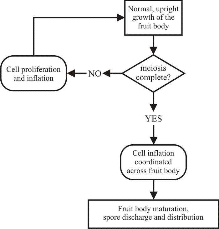 |
| Fig. 25. Simplified flow-chart describing normal upright growth of the mushroom fruit body of Coprinopsis cinerea. Modified after Moore, Greening, Hatton & Novak Frazer, 1994. |
The perception time in Coprinopsis cinerea is 7 minutes (this is the minimum exposure to an altered gravity vector required to cause a reaction). The reaction time in C. cinerea is about 21 minutes; this being the minimum time to elapse after experimental reorientation of the stem before a reaction (in the form of stem curvature) becomes evident. There is no relationship between extent of gravistimulation and rate of response or reaction time. Gravistimulation is not additive, and the angle of bending attained depends on gravistimulation dose (sustained stimulation being required for sustained bending). Perception of the gravity vector does not involve modulation of Ca2+ metabolism, proton channels nor stretch-activated ion channels (Moore, Novak Frazer, Hatton, Greening, Bourne & Robson, 1994). We conclude that the fundamental mechanism might be that gravity perception involves statoliths (possibly nuclei) acting on the actin cytoskeleton and triggering specific vesicle/microvacuole release from the endomembrane system (Moore, Hock, Greening, Kern, Novak Frazer & Monzer, 1996). Most of these observations are incorporated into a gravitropism flow-chart (Figs 25 and 26) (Moore, Greening, Hatton & Novak Frazer, 1994), which also indicates that two signals emanate from the upper regions of the stem, one promotes the process of gravitropic bending, and is followed by a second signal which compensates for excess bending and adjusts the stem apex to the vertical. Although no such growth factors or hormones have so far been isolated from any higher fungus, we have developed a method to extract compounds which cause vertical stems to bend. There appear to be two components which have recognisably different activities in the bioassay. These we call Fungiflex-1 and Fungiflex-2. Though the preparations are still impure, a structure determination seems accessible (Novak Frazer, unpublished observations).
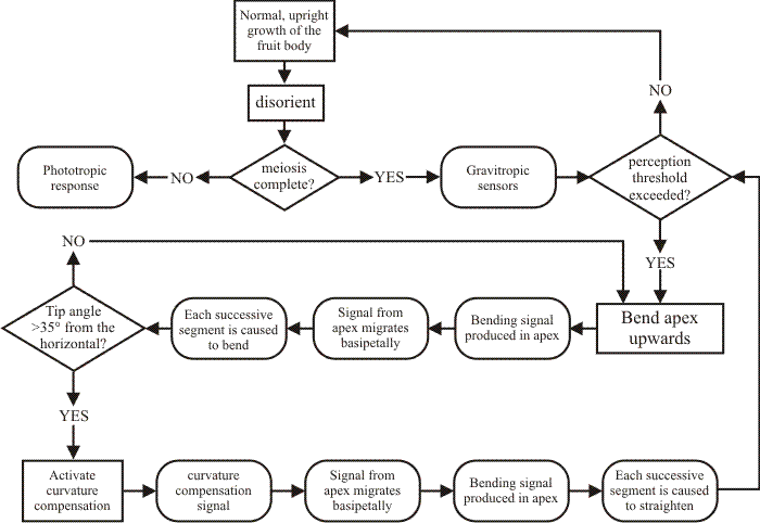 |
| Fig. 26. Flow-chart of the gravitropic response in fruit body stems of Coprinopsis cinerea. Note that formalising the summary in this way emphasises that two signals emanate from the upper regions of the stem; one promotes the process of gravitropic bending, and is followed by a second signal which compensates for excess bending and allows the stem apex to adjust to the vertical. Modified after Moore, Greening, Hatton & Novak Frazer, 1994. |
[TOP]
Updated December 7, 2016