
16.15 Mycoparasitic and fungicolous fungi
In nature, fungi grow in communities of organisms; those communities include microbes, plants, animals and many other fungi. Fungi in the same substrate will compete with one another for control of that nutrient source (and space, Kolesidis et al., 2019) and the competition may be so vigorous that the most successfully competitive fungi specialise in being parasitic on other fungi. After all, production of hydrolytic enzymes into the substrate is the lifestyle that enables saprotrophic fungi to exploit dead substrates, and the same capability enables pathogenic fungi to penetrate their animal, plant or even fungal hosts.
Many fungi are able to secrete glucanases and chitinases, which give them the ability to attack living hyphae of filamentous fungi, causing wall-degradation and consequent autolysis, and there are many species of fungi which are parasites of other fungi. However, fungi growing with or on other fungi can have many different sorts of relationships, varying from indifferent, saprotrophic, commensal, symbiotic, or a range of intensities of parasitic, and it is not always clear which relationship applies.
Before we go into further detail we should emphasise that some, perhaps many, fungi are able to parasitise organisms from different kingdoms; for example, Fig. 13 shows Arthrobotrys oligospora (Ascomycota) acting as a mycoparasite and invading the epidermis of barley roots. Colonisation of plant roots by Arthrobotrys is believed to be as an endophyte, though it may aid the plant host by making the roots more resistant to plant parasitic nematodes and/or fungal pathogens. We first encountered Arthrobotrys in its role as a nematode-trapping fungus (see Fig. 7, Chapter 15; CLICK HERE to view the page), so this species is capable of penetrating the tissues of all three eukaryotic kingdoms (Nordbring-Hertz, 2004).
|
Fig. 13. Versatility in a fungal pathogen able to
attack all three eukaryotic kingdoms. Arthrobotrys oligospora
is a prime example of a nematode-trapping fungus but here, in A,
it is shown as a mycoparasite coiling around a hypha of Rhizoctonia
solani. An unaffected hypha of Rhizoctonia is shown to the
left of the image; scale bar = 10 µm. B shows early
colonisation of the epidermis of barley roots by A. oligospora.
See also the nematode-trapping
devices of Arthrobotrys oligospora shown in Fig. 6 in Chapter
15. Bar = 20 µm. From Nordbring-Hertz, 2004; reproduced with permission
from Elsevier.
|

Another example occurs in the genus Metarhizium, some species of which are host-specific, like the specific locust pathogen Metarhizium acridum, whilst the broad host range Metarhizium robertsii, which is an insect pathogen, also colonises plant roots endophytically, and may even be a plant symbiont. Functional genomic approaches confirmed that Metarhizium robertsii up-regulates different genes in the presence of plants and insects, demonstrating that it has specialist genes for each side of its bifunctional lifestyle. In addition, both Metarhizium species secreted more numerous proteins than were secreted by host-specific plant pathogens or non-pathogenic fungi. This implies that in the greater complexity of interactions between Metarhizium species and their environments there may be more associations to be found (St. Leger et al., 2011); certainly, although fungi are major drivers of ecosystem health, they are strongly influenced by environment, and their interactions with bacteria (Araldi-Brondolo et al., 2017).
Various genera of entomopathogenic fungi have displayed the ability to colonise a wide variety of plant species in different families, both naturally and artificially following inoculation, and then confer protection against fungal plant pathogens as well as insect pests. Consequently, there is great research interest now in the possibility of using entomopathogenic fungi as endophytes for dual biological control of both fungal and insect pests (Jaber & Ownley, 2018).
The term mycoparasitism refers specifically to parasitism of one fungus (the host) by another (the mycoparasite). The word hyperparasitism has been used by mycologists for the same phenomenon, but originated as a description of insects that parasitise other parasitic insects, and is not really appropriate unless the host is itself a parasite. Mycoparasitism describes relationships in which one living fungus acts directly as a nutrient source for another; there is another term, fungicolous, which is used for those relationships (in the majority) in which nutrient transfer directly from live host to live parasite has not been demonstrated. Fungicolous fungi have a constant association with another fungus, but the exact nature of that relationship is difficult to establish (Jeffries, 1995).
By analogy with plant pathogens (see the section entitled Necrotrophic and biotrophic pathogens of plants in Chapter 14; CLICK HERE to view page) mycoparasitic relationships (Table 5) can be described as necrotrophic or biotrophic (Karlsson et al., 2017) :
- necrotrophic, which are destructive parasites that invade and kill their hosts; these fungi often grow well as saprotrophs and include many of the Ascomycota that form large numbers of asexual spores (so-called ‘mitosporic genera’), such as species of Gliodadium and, especially, Trichoderma, as well as members of the ‘Zygomycota’ like Dicranophora and Spinellus, and the Oomycete Pythium. Although Pythium is not a fungus, it rather nicely illustrates the specialisation involved in mycoparasitism because the mycoparasitic species require thiamine for growth in vitro, and are unable to utilise inorganic nitrogen sources, these physiological features representing nutritional deficiencies in comparison plant-pathogenic Pythium spp. These have been classified on the basis of the level of interaction between the parasite and its host, which may be through:
- hypha-to-hypha interference (contact necrotrophs) an example being hyphae of Cladosporium, which cause necrosis and death of basidia of the plant-pathogenic fungus Exobasidium camelliae without penetrating the living basidia (Mims, Hanlin & Richardson, 2007).
- penetration of the hyphae of the host by parasitic hyphae (invasive necrotrophs).
- biotrophic, where a balanced relationship is established and the parasite grows on the still-living mycelium of the host fungus. Most biotrophic mycoparasites are members of the traditional Zygomycota, for example the genera Dispira, Dimargaris, Piptocephalis and Tieghemomyces; these can be grown as dual cultures with their hosts, but without the host fungus they grow poorly in vitro. These have been classified on the basis of the exact nature of the intimate relationship between the parasite and its host, or the host-parasite interface as it is called (Table 5), which may be through:
- entry of the complete thallus of the mycoparasite into the host (intracellular biotrophs; mostly chytrids and oomycete organisms that make their way into their host cytoplasm and absorb nutrients directly from it),
- formation of haustoria from the parasitic hyphae that penetrate the host (haustorial biotrophs),
- specialised contact cells which accomplish direct cytoplasmic continuity with the host through fine pores in the hyphal walls forming interhyphal channels (fusion biotroph).
| Table 5. Types of mycoparasite and the nature of the host-parasite interface involved | |
| Type of relationship | Host-parasite interface |
| Contact necrotroph | Fungi in contact; no penetration of host mycelium by parasitic hyphae; cytoplasm of the host degenerates and hyphal lysis may occur. |
| Invasive necrotroph | Fungi in contact (Fig. 13A); hyphae of the parasite penetrate and enter the host; degeneration of host cytoplasm occurs rapidly, often followed by hyphal lysis. |
| Intracellular biotroph | Complete thallus or mycelium of the mycoparasite enters the hypha of the host; cytoplasm of the host remains healthy. |
| Haustorial biotroph | Hypha of the host is penetrated by a short haustorial branch from the hypha of the parasite; host cytoplasm remains healthy. |
| Fusion biotroph | Fungi in intimately close physical contact; parasitic hyphae often coiled around the host hyphae, the two pressed closely together; micropore(s) develop between the closely pressed hyphae, or from a short penetrative hyphal branch of the parasite; host cytoplasm remains healthy. |
| Table modified from Jeffries (1995). | |
Mycoparasitism depends on hyphal recognition and interaction, and many
specific gene-regulation events in both parasite and host; for example, see the
paper by Morissette et al. (2008) about gene expression during the
interaction of the mycoparasite Stachybotrys elegans with its host
Rhizoctonia solani.
There are several sorts of interaction between fungal hyphae, some of which we
have already described in detail. Intraspecific interactions
involve of the same species, though they may come from different mycelia. If the
interacting hyphae come from within a single mycelium hyphal fusions occur
readily to improve the interconnections within the mycelium (see the section
entitled Hyphal fusions and mycelial interconnections in Chapter 5;
CLICK HERE to view the page).
Interactions between different mycelia can involve vegetative compatibility reactions that determine the individuality of mycelia and are able to regulate the degree of anastomosis and heterokaryosis within a hyphal network (see Fig. 7 in Chapter 7 and the associated text; CLICK HERE to view the page). These intraspecific interactions can make the competition between different individual mycelia within a substrate evident when the interaction gives rise to antagonism within the contact zone between opposing mycelia to form, for example, zone lines in infected timber (see Fig. 8 in Chapter 7, CLICK HERE to view the page; and Fig. 3 in Chapter 13, CLICK HERE to view the page). In extreme cases, where one partner in the interaction is extremely dominant, destruction of one mycelium by the dominant one can result in the transfer of nutrients during these intraspecific interactions, so they can become mycoparasitic. There can also be a test for sexual compatibility, as an essential prelude to plasmogamy and karyogamy as part of the sexual reproductive cycle (see the section entitled The process of sexual reproduction (and subsequent sections) in Chapter 8; CLICK HERE to view the page).
Interspecific interactions may initially depend on some of the same mechanisms. They have been characterised as neutralistic, mutualistic or competitive (Cooke & Rayner, 1984; Dighton, White & Oudemans, 2005):
- in neutralistic interactions the hyphae intermingle without any apparent reaction to one another;
- in mutualistic interactions both individuals derive some benefit, which increases the survival value of both;
- competitive interactions are detrimental to both species because the antagonism experienced reduces fitness.
Competitive interactions generally result in one of the competing fungi proving to be more aggressive than the other and the expression of this is that the more aggressive fungus captures more nutrients than the less aggressive. In the broadest sense, the aggressive fungus is acting as a mycoparasite, but this can be a capture of substrate rather than parasitic absorption of nutrients directly from a host, so this is where the term fungicolous is most useful because it covers the full range of associations between fungi that live together whatever the biological nature of the association (Gams, Diederich & Poldmaa, 2004).
The most aggressive fungicolous fungi are the true mycoparasites that are divided into the two groups, necrotrophs and biotrophs (Table 5, above), where the necrotroph is so strongly aggressive that it dominates and ultimately kills its partner/host. Close direct contact between necrotroph and host, often with the necrotrophic hyphae coiled around hyphae of the host (Fig. 13A), helps exchange of nutrients without loss to competing third-party microbes. Biotrophic mycoparasitic relationships are physiologically balanced; the parasite being highly adapted to coexisting with the host for extended periods of time, and often forming specialised infection structures or host-parasite interfaces (Table 5).
Mycoparasites are probably very significant in the natural environment because fungi are major contributors to natural communities and these relationships between fungi must play an important role in development of community structure. Fungicolous fungi are common, widespread, and numerous. Many fungi occur in nature in obligate associations with the mycelium or sporing structures of other fungi; these may be parasitic relationships; they are certainly interdependent relationships. DNA metabarcoding has revealed complex communities of fungicolous fungi, largely dominated by Ascomycota, within fungal sporocarps of wood-decay fungi collected in a forest in northeast Finland (Maurice et al., 2021). Host species was the major determinant of community composition and diversity of fungicolous fungi, and the mean species diversity was consistently higher in short-lived and resupinate sporocarps compared to long-lived and pileate ones, perhaps due to a more hostile environment for fungal growth in the latter.
Because it is often difficult to obtain clear evidence of mycoparasitism it is frequently inferred as the lifestyle of a fungus for the reason that the mycoparasite causes distinctive growth inhibitions and/or abnormalities in the hyphae of the alleged host. Overall this makes it difficult to establish the exact number of fungi known to grow on other fungi (Jeffries, 1995). But there are estimates, and the estimated numbers are large. The 10th edition of the Dictionary of the Fungi (Kirk et al., 2008) puts the numbers at 1,100 species of fungi parasitising 2,500 other species of fungi, with probably at least a further 2,000 species of fungi, of all groups, growing specifically on lichens (lichenicolous fungi). Although these numbers are large they are minima; fungicolous fungi occur in all habitats and it is probable that mycoparasitism occurs more widely than currently imagined. After all, because fungi are such common, even dominant, members of natural populations in all habitats, the ability to harvest nutrients from either their living or dead hyphae must have very positive selective potential.
Jeffries (1995) details two specific examples of mycoparasitic relationships:
- necrotrophic invasion of spores of arbuscular mycorrhizal fungi. The genera Acaulospora, Glomus, Gigaspora, Scutellospora, and Sclerocystis (Glomeromycota) are the characteristic and ubiquitous arbuscular mycorrhizal fungi. The spores of some of these are the largest known in the fungal kingdom and they can be extracted easily from field soils by simple sieving. In any sample of spores extracted like this there is usually a proportion, often the majority that are parasitised; the spore wall being perforated by many fine radial canals caused by penetration of the wall by hyphae of mycoparasitic fungi, or by rhizoids as parasitism of these spores by chytrids is probably widespread in wet soils. Members of the Oomycota, including Spizellomyces and Pythium-likefungi are also frequently seen within arbuscular mycorrhizal spores. Other fungi can often be isolated easily from arbuscular mycorrhizal spores and seem to be facultative parasites, being essentially saprotrophs with some ability to attack live spores. One study of mycoparasites of spores of Gigaspora gigantea reported the recovery of 44 species of fungi from surface-disinfected spores; Acremonium spp., Chrysosporium parvum, Exophiala werneckii, Trichoderma spp., and Verticillium sp. (all Ascomycota) were most frequently isolated from healthy spores, while Fusarium spp., Gliomastix spp.(both Ascomycota) and Mortierella ramanniana (Mortierellomycotina) were typically isolated from dead or dying spores. Several examples of mycoparasitism of arbuscular mycorrhizas by various aquatic fungi have been described from specimens of the Lower Devonian Rhynie chert; demonstrating the presence of mycoparasitism in a 400-million-year-old ecosystem (Taylor et al., 2015; Krings et al., 2018) .
- biotrophic invasion of mucoralean hosts by haustorial mycoparasites. Mycoparasites that penetrate their host with haustoria are highly specialised and frequently unable to grow in the absence of the host. Examples, all from the zygomycetes, are parasitic species of Piptocephalis (Zoopagomycotina) and Dimargaris (Kickxellomycotina) growing on living saprotrophic hosts from the Mucoromycotina (e.g. Pilobolus, Pilaira and Phycomyces) on the dung of herbivorous animals, especially rabbit pellets. Piptocephalis species are also extremely common in many woodland and pasture soils, from which they can be isolated using a baiting technique (spreading the soil sample on a medium already colonised by the host fungus).
These examples involve interactions on a microscopic scale. The impact of parasitic fungi is most obvious, however, in examples where the parasitic fungus produces a macroscopic fruit body from a macroscopic fruit body of its host. Fig. 14 shows the example of Boletus parasiticus (also known as Xerocomus parasiticus) growing on a fruit body of Scleroderma citrinum.
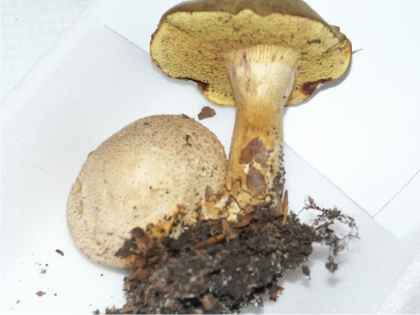 |
| Fig. 14. Mycoparasitic macrofungi. A fruit body of Boletus parasiticus (also known as Xerocomus parasiticus) growing on a fruit body of the Common earthball, Scleroderma citrinum. Collected in Harlow Carr Gardens, Harrogate in 2005. Photograph by David Moore. |
This bolete species is said to be edible, but its host is poisonous, so it is not recommended for eating! The host, Scleroderma citrinum, is mycorrhizal with hardwoods and conifers; it is common and frequently found on the roots of trees growing in mossy, boggy areas in late summer and autumn across the North Temperate zone. Although the parasite is described in field guides as ‘rare’ its host is so common that the parasitism is encountered regularly. There are many other examples of parasitism amongst our common field fungi (see Michael Kuo’s Key to Mycotrophs on his MushroomExpert.Com Web site at: http://www.mushroomexpert.com/mycotrophs.html).
Such mycotrophism is not always as obvious as it is in this specimen of Boletus parasiticus (Fig. 16); it can go unnoticed because the host mushroom (or other fruit body) can be highly reduced, blackened and almost unrecognisable, or the host may be underground. For example, Squamanita odorata is an agaric which is parasitic on other agaric fruit bodies, but because the host tissues are reduced to unrecognisable galls, molecular techniques were necessary to identify that the host is Hebeloma mesophaeurn (Mondiet, Dubois & Selosse, 2007). Not all of the best examples are basidiomycete ‘mushrooms and toadstools’. For example, the ascostromata of the club fungus Cordyceps (or Tolypocladium; Ascomycota) are often found growing out of the subterranean fruit bodies of the false truffle Elaphomyces (Ascomycota): see the illustrations hyperlinked in the Resources Box). We are more familiar with Cordyceps (caterpillar fungus) as a widespread parasite of insects (e.g. Cordyceps militaris common throughout the Northern Hemisphere as a parasite of the larvae and pupae of butterflies and moths) so this is another reminder that parasitic genera can have species that specialise on hosts from different kingdoms (compare Fig 13). The Himalayan caterpillar fungus (Ophiocordyceps sinensis) has become one of the world’s most valuable biological commodities, high-quality pieces selling for more than three times their weight in gold, because of the high value placed on these fungi in traditional Chinese medicine. Inevitably, overharvesting of this fungus by collectors is affecting the ecosystem of the alpine regions of Himalaya threatening many endangered plant and animal species of that area (Hopping et al., 2018: Roy, 2021).
Resources Box Ascostromata of the club fungus Cordyceps (Ascomycota) are often found growing out of the subterranean fruit bodies of the false truffle Elaphomyces (Ascomycota). View the following web pages for illustrations: Michael Kuo’s Key to 25 mushroom-eating mushrooms and fungi (mycotrophs) on his MushroomExpert.Com website [URL: http://www.mushroomexpert.com/mycotrophs.html]. Tom Volk’s Fungi website [URL: http://botit.botany.wisc.edu/toms_fungi/jan98.html]. |
It is not known when mycoparasitism evolved, though we have noted above the occurrence of mycoparasites of arbuscular mycorrhizas in 400-million-year-old fossils in the Rhynie chert. Macrofungi are not good candidates for fossilisation, but mycoparasitism of an agaric mushroom in Early Cretaceous (100 million-year-old) amber has been demonstrated (Poinar & Buckley, 2007), this being the oldest known fossil of an agaric mushroom. Interestingly, in this specimen the mycoparasite was itself infected by a hypermycoparasite; so these observations also show that complex patterns of mycoparasitism were well developed in the Cretaceous.
In the present day a large number of fungi parasitise others of their kind and those that attack commercially grown mushrooms often cause major economic losses to the industry; so as our final example we will briefly describe some of the mycoparasites that cause disease in mushroom farms, particularly those of the most widely cultivated mushroom, Agaricus bisporus. Cultivated mushrooms are prone to bacterial and virus diseases that can cause significant crop losses, but these are outside our current area of interest. The two most common and geographically most widespread mycoparasites of A. bisporus are the Ascomycota Mycogone perniciosa (cause of the disease known as wet bubble or white mould) and Verticillium fungicola (cause of the disease known as dry bubble or brown spot). These parasites can cause serious economic losses to the mushroom farmer, and they also cause similar diseases of the Paddy Straw Mushroom, Volvariella volvacea, in cultivation.
Vegetative mushroom mycelium is not adversely affected by M. perniciosa in vitro until after formation of the strands from which the fruit bodies develop. Verticillium hyphae grow over the mushroom mycelium in vitro, eventually causing severe necrosis of the Agaricus mycelium. Verticillium causes more mushroom necrosis than M. perniciosa and it also kills mushroom mycelium, whereas M. perniciosa does not. When M. perniciosa grows on a mushroom fruit body its growth is thick, velvety and white (which is why it is called white mould), while Verticillium produces a fine, greyish-brown felted growth (which is why it is sometimes called a cobweb mould though this is usually applied to a Cladobotryum infection; see below).
A common result of parasitism by M. perniciosa and Verticillium is a drastic effect on mushroom development, and mushroom abnormalities are the prime disease symptoms. The extent of the symptoms depends on the stage of development reached at the time of infection; the earlier the infection occurs, the greater the deformity caused. The most extreme effect is for a spheroidal mass of undifferentiated tissue to be formed rather than a mushroom; these are the ‘bubbles’ of these two bubble diseases. The undifferentiated masses caused by M. perniciosa can be 5 cm or more across, whereas those caused by Verticillium are usually less than 1 cm in diameter.
Infection with Verticillium at later stages of development causes developmental deformities, including bulbous stems with vestigial caps and fruit bodies in which the tissues are broken and peeled back (Largeteau & Savoie, 2008). Infection of well-developed fruit bodies by M. perniciosa commonly results in abnormal enlargement of the gills (lamellae) as they become covered by the parasite. Internally, Mycogone-infected mushrooms become wet and develop a foetid odour, and drops of a clear amber coloured liquid are often extruded from the mushroom (which is why it is called wet bubble disease). Verticillium-infected sporophores are dry and shrivelled at first (= dry bubble disease), but in both cases bacteria invade and a bacterial rot results. Although these mycoparasites are certainly able to produce enzymes capable of degrading mushroom hyphal wall polymers most of the rot that occurs in the final stages of these infections is due to secondary (opportunistic) invasion of the necrotic tissues by bacteria and other fungi.
Macrofungi in the wild, particularly agarics, are commonly parasitised by members of the genus Cladobotryum (Ascomycota), and C. dendroides is also a common pathogen of cultivated mushrooms. The parasite causes general decomposition of the mushroom tissues, characteristic deformities do not occur. C. dendroides growth over the surface of mushroom beds is conspicuous, giving rise to the common name ‘cobweb’. Mushrooms engulfed by the spreading mycelium of the parasite turning pale brown and develop a soft rot, particularly at the base of the stem. Mushroom crops can be damaged by a range of saprotrophic fungi, especially those that favour lignocellulosic substrates, which may be present in poorly-prepared composts or timber containers. These are usually considered to be weed fungi rather than parasites.
Generally speaking, many cultivated fungi are not very vigorous (the Pleurotus spp. that are cultivated may be an exception to this generalisation), so they tend to come second in any competition for the substrate. Schizophyllum, Stereum, Coriolus, Coniophora, Merulius (all Basidiomycota) and Hypoxylon (Ascomycota) are among the genera of wood-destroying fungi that have been recorded in mushroom farms using timber containers or in traditional outdoor log cultivation of Lentinula edodes (shiitake). The most common weeds on farms that use compost substrates are the smaller ink-cap mushrooms, such as Coprinopsis cinerea and its relatives, and the extremely common and widespread Trichoderma (Ascomycota). Trichoderma pleurotum and Trichoderma pleuroticola are highly aggressive parasites causing green mould diseases of the oyster mushroom Pleurotus ostreatus that threaten commercial production of oyster mushrooms around the world and may be a major infection risk for other mushroom farms (Komon-Zelazowska et al., 2007).
Trichoderma spp. are common in most soils; they are competitive saprotrophs, opportunistic parasites of other fungi, and possibly symbionts of plants (Harman et al., 2004). Trichoderma viride parasitises Rhizoctonia solani, which causes the disease of seedlings known as ‘damping-off’, and Armillaria and Armillariella, which are important pathogens of trees. At least some strains of Trichoderma establish vigorous and long-lasting colonisations of root surfaces by penetrating the epidermis and a few cell layers beneath. This is far from parasitic because the fungus enhances root growth and development and the uptake and use of nutrients, and induces resistance responses in the plant that protect it from several plant pathogens as well as abiotic stresses. A very high proportion of agricultural crop loss is due to diseases caused by soil microbes and strains of Trichoderma (and its relative Gliocladium) have been developed that can be used for biocontrol of crop pathogens; in other words Trichoderma is being used as a mycopesticide. Trichoderma, though, is a serious contaminant in the production of mushrooms of the genus Pleurotus, which very often produces large crop losses. So, it is interesting to note that acetone-extracts of dried fruit bodies of the polypore Pycnoporus inhibited growth of Trichoderma strains (isolated from infected substrate in Pleurotus production modules) in both agar and wheat straw test cultures (Talavera-Ortiz et al., 2020).
Another example is Scytalidium parasiticum (Ascomycota; anamorph of Xylogone species), which has been reported as a potentially useful necrotrophic mycoparasite of Ganoderma boninense causing basal stem rot of oil palm in Malaysia and elsewhere. The mycoparasite was isolated from Ganoderma-infected oil palms in plantations and then demonstrated to be environmentally-friendly in the sense of having low to very low toxicity to hamster cell lines in vitro, and nontoxic when fed to rats. Scytalidium parasiticum is therefore seen as a potential candidate for biocontrol of basal stem rot in oil palm caused by Ganoderma boninense, there being no other effective treatments (Goh et al., 2016). We will describe more general aspects of the use of parasitic fungi as mycopesticides in more detail in Chapter 17.
Updated May, 2021