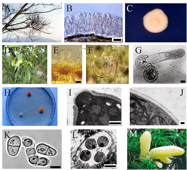
Macro- and micromorphology showing the diversity of the Taphrinomycotina.
|
|
Macro- and micromorphology showing the diversity of representative taxa in the Taphrinomycotina. A, Witches’ broom disease of a Japanese cherry tree (Cerasus yedoensis) caused by Taphrina wiesneri. B, Mature asci of T. wiesneri on leaf tissue of a Japanese cherry tree showing ascospore budding (blastoconidium formation). C, A colony on potato dextrose agar. D, Symptoms of peach leaf curl disease caused by Taphrina deformans. E, Hymenium of Taphrina caerulescens, causal agent of oak leaf curl. F, Galls induced by Protomyces inouyei on stem of Youngia japonica. G, Germination of a thick-walled resting spore of P. inouyei in water. H, Colonies of Rhodosporidium toruloides (anamorph: Rhodotorula glutinis) (top), Saitoella complicata (right) and Taphrina wiesneri (left) on potato dextrose agar. I, Saitoella complicata: TEM showing enteroblastic budding, characteristic of basidiomycetous yeast. J, Saitoella complicata. TEM showing the cell wall ultrastructure of the ascomycete type composed of one thin, dark layer and a broad, light inner layer. K, Schizosaccharomyces pombe. Fission and an ascus containing four ascospores. L, Pneumocystis jirovecii. Mature cyst containing intracystic bodies (endospores). M, Neolecta vitellina. Bright yellow fruit-bodies that can grow several cm tall. Scale bars: B, E, G = 20 μm; I = 0.5 μm; J = 0.1 μm; K = 5 μm; L = 1 μm. Modified from Sugiyama et al. (2006) using graphic files kindly supplied by Prof. Junta Sugiyama, TechnoSuruga Co. Ltd., Tokyo, Japan. Reprinted with permission from Mycologia. © The Mycological Society of America. |
Close the window to return to 21st Century Guidebook to Fungi
This is a Resources Box from the 21st Century Guidebook to Fungi:© David Moore, Geoffrey D. Robson and Anthony P. J. Trinci 2019