Some characteristic morphological features of glomeromycotan fungi
|
|
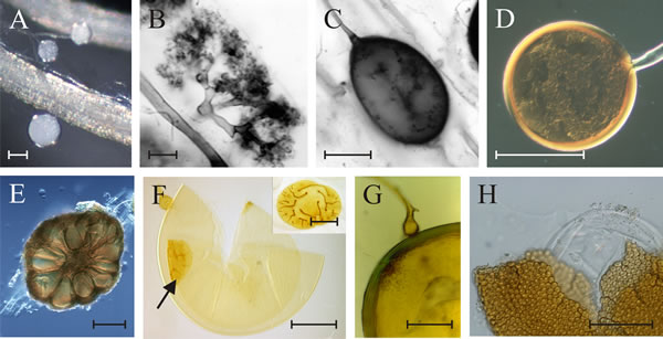
Some characteristic morphological features of fungi belonging to the subphylum Glomeromycotina. A, roots of Plantago media colonised with hyphae and spores of Glomus clarum. B, arbuscule of Glomus mosseae stained with chlorazol black. C, Vesicle of Glomus mosseae. D, Spore of Glomus sp. showing the hyphal attachment. E, Section of a sporocarp of Glomus sinuosum with spores grouped around a hyphal plexus and covered by a layer of hyphae. F, Spore of Scutellospora cerradensis, showing bulbous sporogenous cell and inner flexible walls with germination shield (arrow); inset: germination shield of S. scutata in face view (photomicrographs by Fritz Oehl, Agroscope Reckenholz-Tänikon, Switzerland). G, Germinating spore of Gigaspora decipiens with sporogenous cell, warty germination layer and germination hypha. H. Spore of Acaulospora denticulata with tooth-like wall ornamentations and inner germinal walls. Scale bars = 100 µm (A, E, F, G), 50 µm (D, H), 5 µm (B, C). Modified from Redecker & Raab, 2006 using graphic files kindly supplied by Prof. Dirk Redecker, Université de Bourgogne, France. Reprinted with permission from Mycologia. © The Mycological Society of America. |
Close the window to return to 21st Century Guidebook to Fungi
This is a Resources Box from the 21st Century Guidebook to Fungi:© David Moore, Geoffrey D. Robson and Anthony P. J. Trinci 2019