Light micrographs of representative chytrids
|
|
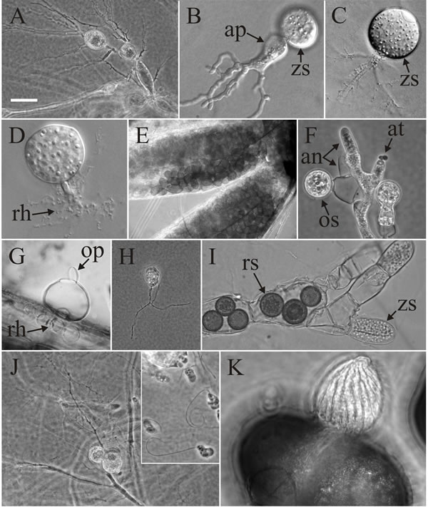
Light micrographs of representative chytrids to give some indication of their biodiversity. A, Catenomyces persicinus polycentric thallus with apophysate (swollen) rhizoidal axis and intercalary zoosporangia. B, Spizellomyces plurigibbosus monocentric, inoperculate zoosporangium (zs), with swollen, apophysate rhizoidal axis (ap) and branched rhizoids that are blunt at the tips. C, Chytriomyces hyalinus monocentric, operculate zoosporangium (zs) with long branched rhizoids that taper to less than 0.5 µm at the tips. D. Terramyces subangulosum monocentric, inoperculate sporangium with a thick rhizoidal axis, and densely branched rhizoids (rh) that taper to less than 0.5 µm at tips. E, Coelomomyces stegomyiae elliptical resting spores inside the anal gills of its mosquito host. F, Monoblepharis polymorpha mature zygote or oospore (os), empty and mature antheridia (an) and male gametes (antherozoids, at) emerging from antheridium. G, Catenochytridium sp., monocentric, operculate (op) zoosporangium with catenulate (= chain-like) rhizoids (rh). H, Lobulomyces angularis monocentric, operculate sporangium with thread-like rhizoids that branch several µm from the sporangial base. I, Rozella allomycis parasitising hyphae of Allomyces (another chytrid, though now placed in the related phylum Blastocladiomycota). The parasite grows inside the host and causes it to produce hypertrophied, highly septate cells within which the parasite forms thick-walled resting spores (rs) or unwalled zoosporangia (zs) that use the host’s cell wall as its own. J, the polycentric thallus of Polychytrium aggregatum with at least two sporangia; the inset shows the zoospores of P. aggregatum. K. Blyttiomyces helicus growing on pollen grain and forming an epibiotic, inoperculate sporangium with distinct helical pattern. Scale: approximate sizes indicated by scale bar in A = 10 µm. Images by Dr Timothy Y. James, Department of Ecology and Evolutionary Biology, University of Michigan, Dr Joyce E. Longcore, School of Biology and Ecology, The University of Maine and Dr Peter M. Letcher, Department of Biological Sciences, University of Alabama. Figure modified from James et al. (2006) using graphic files kindly supplied by Dr T. Y. James. Reprinted with permission from Mycologia. © The Mycological Society of America. |
Close the window to return to 21st Century Guidebook to Fungi
This is a Resources Box from the 21st Century Guidebook to Fungi:© David Moore, Geoffrey D. Robson and Anthony P. J. Trinci 2019