Ascomycete yeasts in culture and in scanning electron micrographs. |
|
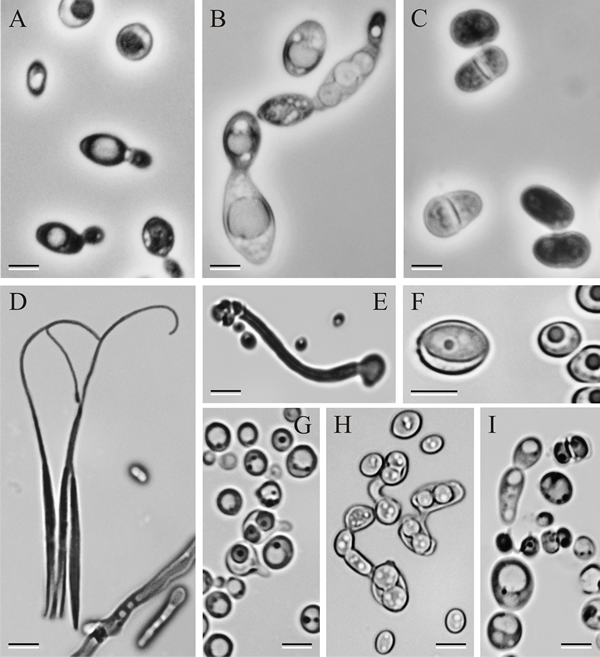
Ascomycete yeasts in pure culture. A, Saccharomyces cerevisiae. Cells dividing by multilateral budding. B, Saccharomycodes ludwigii. Cell division by bipolar budding. Note the ascus with four spherical ascospores above the dividing cell. C, Schizosaccharomyces pombe. Cell division by fission. Once divided, the newly formed cells often will be morphologically indistinguishable from cells formed by budding. D, Eremothecium (Nematospora) coryli. Free, needle-shaped ascospores with whip-like tails of extended wall material. Members of this group are some of the few yeasts that are plant pathogens. E, Pachysolen tannophilus. A single ascus forms on the tip of an elongated refractile tube. The ascus wall becomes deliquescent and releases four hat-shaped ascospores. P. tannophilus was the first yeast discovered to ferment the pentose sugar D-xylose, a major component of hemicellulose from biomass. F, Lodderomyces elongisporus. Persistent ascus with a single ellipsoidal ascospore. This is the only species of the clade, that includes Candida albicans and C. tropicalis, which is known to form ascospores. G, Torulaspora delbrueckii. Asci with 1–2 spherical ascospores. Asci often form an elongated extension that may function as a bud conjugant. H, Zygosaccharomyces bailii. Asci with spherical ascospores. Often there are two ascospores per conjugant giving rise to the term ‘dumbbell-shaped’ asci. This species is one of the most aggressive food spoilage yeasts known. I, Pichia bispora. Ascospores are hat-shaped and released from the asci at maturity. Hat-shaped ascospores are produced by species in a variety of different genera (see Fig. 3.12A, for example). Figs 3.11A–C, phase contrast. Figs 3.11D–I, bright field; all photographs by Dr C. P. Kurtzman, ARS, USDA. Scale bars = 5 µm. Modified from Suh et al. (2006) using graphic files kindly supplied by Dr Sung-Oui Suh, American Type Culture Collection, Mycology Collection. Reprinted with permission from Mycologia. © The Mycological Society of America.
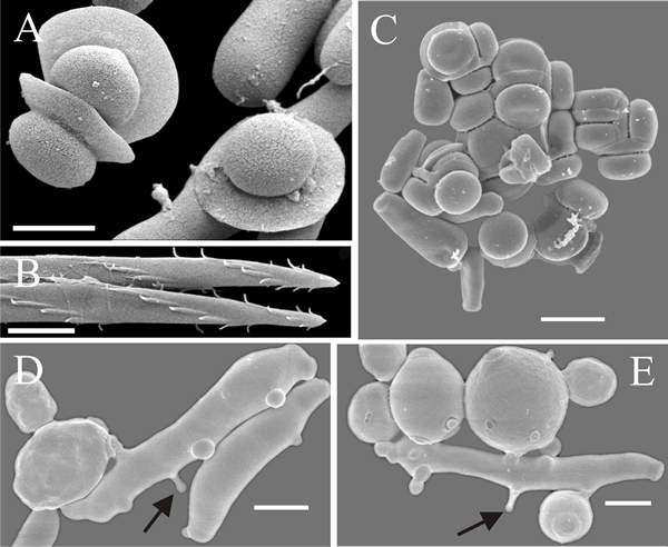
Scanning electron micrographs of yeasts. A, Ascospores of Kodamaea anthophila. B. End of ascospores of Metschnikowia borealis released by treatment of the ascus with Mureinase. C. Agglutinated ascospores of Saccharomycopsis synnaedendrus. D, E. Elongated predaceous cells of Arthroascus schoenii penetrating ovoid cells of Saccharomyces cerevisiae by means of narrow infection pegs (arrows). Scale bars = 2 µm. Photographs by Dr M.-A. Lachance, Western Ontario University, Canada. Modified from Suh et al. (2006) using graphic files kindly supplied by Dr Sung-Oui Suh, American Type Culture Collection, Mycology Collection. Reprinted with permission from Mycologia. © The Mycological Society of America. |
Close the window to return to 21st Century Guidebook to Fungi
This is a Resources Box from the 21st Century Guidebook to Fungi:© David Moore, Geoffrey D. Robson and Anthony P. J. Trinci 2019